
A team of researchers from San Diego State University (SDSU) and the University of Washington (UW) are collaborating to develop a novel wireless device to record and stimulate brain activity, one of the long-term goals of the Center for Sensorimotor Neural Engineering.
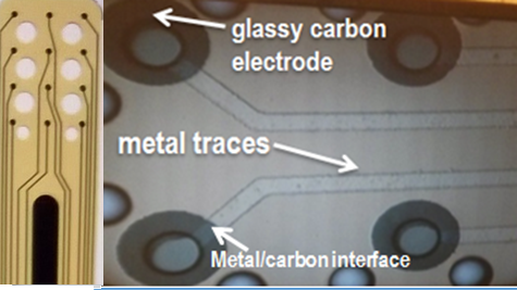
SDSU researchers have created flexible glassy carbon electrodes, now commonly used in a variety of applications, which can be implanted on the surface of the brain to record signals or stimulate specific sites.
Maria Vomero, SDSU graduate student in the NeuroMEMS Lab, said the team “heats the polymer at different temperatures and different rates of speed. We noticed that if we change the parameters, we get a new material every time.”
 Heating the material at high temperatures gets rid of other non-carbon elements, said Scott Seidman, also a graduate student in the NeuroMEMS Lab. “All that’s left is the glassy carbon that fuses together,” he said.
MEMS stands for Microelectromechanical Systems. The NeuroMEMS Lab is led by Associate Professor Sam Kassegne.
After changing the makeup of the material, the research team then mounts the glassy carbon electrodes on a flexible base. One of the benefits of using carbon is the lower corrosion rate, compared with existing metal electrodes.
Vomero said there are probably less than 10 groups around the world working with carbon in this way. She’s been working on this project for the last two years.
Seidman has been focused on what to do with the electrodes after the manufacturing process. “I’m exploring how we minimize ‘noise’ and affect the signal when it is transferred,” he said. “Eventually, the end goal would be to take a signal in your brain, wirelessly send it where it needs to go and use that signal without having to process or encode it,” Seidman said.
One of the systems being developed, named PeSKa—using initials of the collaborators—will be used in electrocorticography (ECoG), which involves surgically implanting a number of electrodes on the surface of the brain (under the skull, but not penetrating the brain tissue). ECoG is regularly performed on people who have epilepsy prior to undergoing surgery to help control seizures. These electrodes record the activity of the brain, allowing researchers to learn more about what brain signals are activated when a person lifts an arm or shakes someone’s hand.
Designing the electrodes so that they will be on the surface of the brain will, in theory, create less damage to the tissue and prevent a foreign body response. Existing electrodes and implanted systems are typically only useful for around eight weeks due to the buildup of scar tissue, Vomero said. Researchers then cannot record brain signals once the scar tissue has built up. By using the glassy carbon electrodes, researchers believe that they can extend the amount of time that they can study brain signals and eventually stimulate certain sites, too.
Vomero, Seidman and SDSU scientists are working on ways to clean the corrosion off of the devices wirelessly in the future. They are also exploring the use of these devices in deep brain stimulation, which is currently FDA-approved for Parkinson’s disease, essential tremor, dystonia and Tourette’s syndrome.
The team is testing the new devices in vivo.
SDSU researchers have filed for a patent to protect the fabrication approach they’ve described above.